A 25-year-old male with a history of multiple episodes of palpitation and near-syncope. What is abnormal?
- Anterior wall myocardial infarction
- Hyperkalemia
- Right bundle branch block
- Wolff-Parkinson-White (WPW) pre-excitation pattern
- Left ventricular hypertrophy
The right answer is:
4. Wolff-Parkinson-White (WPW) pre-excitation pattern
Explanation
Normally the electrical stimulus travels to the ventricles from the atria via the atrioventricular (AV) junction. The physiologic lag of conduction through the AV junction results in the normal PR interval of 0.12 to 0.2 sec. Consider the consequences of having an extra pathway between the atria and ventricles that would bypass the AV junction and pre-excite the ventricles. This situation is exactly what occurs with the WPW pattern: an atrioventricular bypass tract or accessory pathway (AP) connects the atria and ventricles, circumventing the AV junction.
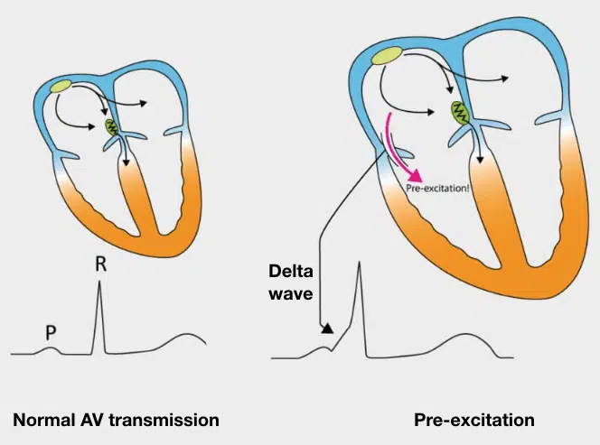
Figure 1: In a small percentage of people, an accessory fiber (atrioventricular bypass tract) connects the atria and ventricles.
Preexcitation of the ventricles with the classic WPW pattern produces the following characteristic triad of findings on the ECG (Figure 2):
- The QRS complex is widened, giving the superficial appearance of a bundle branch block pattern. However, the wide QRS is caused not by a delay in ventricular depolarization but by early stimulation of the ventricles. The T wave is also usually opposite in polarity to the wide QRS in any lead, similar to what is seen with bundle branch blocks (“secondary T wave inversions”).
- BECAUSE OF THE VENTRICULAR PREEXCITATION, the PR interval is shortened (often but not always to less than 0.12 sec).
- The upstroke of the QRS complex is slurred or notched. This is called a delta wave.
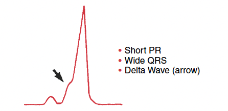
Figure 2: Preexcitation via the bypass tract in the Wolff- Parkinson-White (WPW) pattern is associated with a triad of findings.
Notes on terminology: Use the term “WPW syndrome” to apply to patients with the “WPW pattern” who also have arrhythmias related to the bypass tract.
The WPW "pattern" or Pre-excitation pattern are usually used interchangeably, consisting of the aforementioned triad. This "pattern" can be accidentally found on routine ECG done for other purposes in asymptomatic individuals. However, the term WPW "syndrome" or pre-excitation "syndrome" implies finding the pattern on ECG and the occurrence of tachyarrhythmia episodes.
Clinical Significance:
The classic WPW appearance on ECG has been reported in roughly 1 to 2 per 1000 individuals. In some instances, a familial occurrence is observed. Right ventricular bypass tracts are sometimes associated with other forms of congenital heart disease, especially Ebstein’s anomaly of the tricuspid valve. Left-sided bypass tracts usually are not associated with structural heart disease. The major significance of WPW preexcitation is twofold:
- Individuals with this pattern are prone to arrhythmias, especially paroxysmal supraventricular tachycardia and atrial fibrillation. In the latter instance, if the rate becomes extremely fast, atrial fibrillation may lead to ventricular fibrillation with sudden cardiac arrest. Fortunately, this occurrence is very rare.
- The WPW ECG is often mistaken for either a bundle branch block, due to the wide QRS, or for an MI, due to the negative delta waves or abnormal repolarization (ST-T changes) as in figure 3.
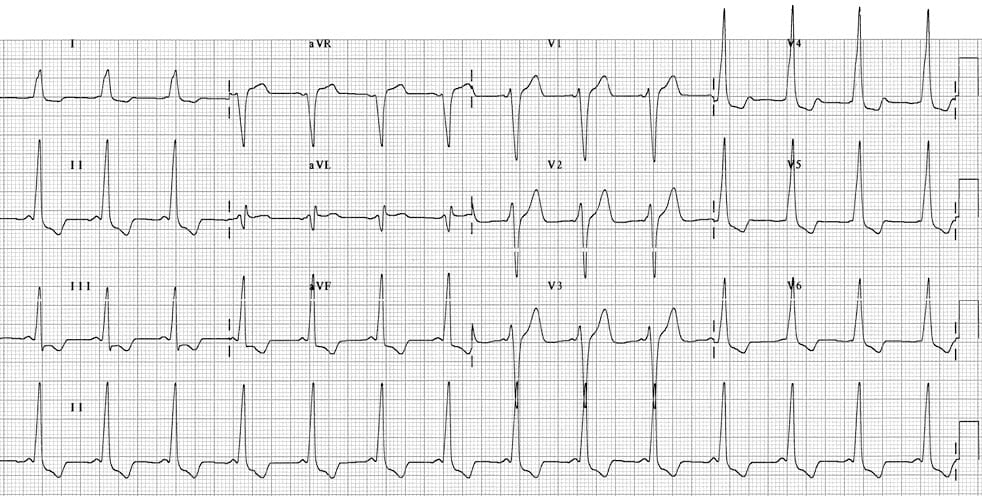
Figure 3:
- Sinus rhythm with very short PR interval (< 120 ms).
- Broad QRS complexes with a slurred upstroke to the QRS complexes — the delta wave.
- Dominant S wave in V1 indicates a right-sided AP — sometimes referred to as “Type B” WPW.
- Tall R waves and inverted T waves in the inferior leads and V4-6 mimic the appearance of left ventricular hypertrophy (LVH) — this is due to WPW and does not necessarily indicate underlying LVH.
References:
- Nathanson LA, McClennen S, Safran C, Goldberger AL. ECG Wave-Maven: Self-Assessment Program for Students and Clinicians. http://ecg.bidmc.harvard.edu.
- Goldberger, Ary L., Zachary D. Goldberger, and Alexei Shvilkin. Clinical electrocardiography: a simplified approach e-book. Elsevier Health Sciences, 2017.
- Buttner R. and Burns E. Life in the fast lane. https://litfl.com/pre-excitation-syndromes-ecg-library/